|
Optical
Coherence Tomography (OCT)
As of November this year we
will be acquiring a special piece of equipment called an
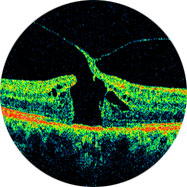
Optical Coherence Tomographer (OCT). This machine
is capable of providing us with a very high resolution
three-dimensional digital scan of the inside of your
eye, much like an MRI scanner at the hospital.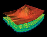
The
scan the OCT provides is so detailed that it can pick up
the very earliest signs of glaucoma – damage to
the microscopic nerve fibres at the back of your eye.
This makes it possible to pick up signs of glaucoma
before it affects your sight, and indeed before it is
detectable using more traditional techniques. This makes
OCT the definitive screening tool currently available
for this condition.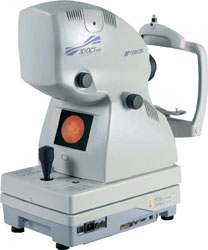
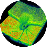
The OCT
also provides us with highly useful information about
other parts of your eye. For example, it is capable of
detecting small areas of swelling that can occur at the
back of your eye in macular degeneration, as well
as any small retinal holes, tears or cysts that may be
present.
We recommend OCT for all of our patients as part of an
ongoing ocular health assessment, but especially for
those people who are most at risk of eye conditions such
as glaucoma and macular degeneration (e.g. individuals
with a family history of eye disease, and/or over the
age of 40). As one would expect, performing an OCT scan
goes above and beyond what is covered by the NHS for a
standard sight test. As a result there is an additional
charge for having this procedure done. Feel free to ask
your optometrist about OCT at your next appointment with
us.
|
|
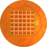
Glaucoma
The scan the OCT provides is
so detailed that it can pick up the very earliest signs
of glaucoma – damage to the microscopic nerve
fibres at the back of your eye. This makes it possible
to pick up signs of glaucoma before it affects your
sight, and indeed before it is detectable using more
traditional techniques. This makes OCT the definitive
screening tool currently available for this condition.
|
Vitreous Detachments
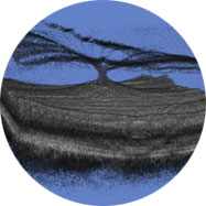
Vitreomacular traction
can clearly be diagnosed through OCT providing
invaluable information as to the current relationship
between the vitreous and the retinal surface. As we get
older the vitreous, the jelly that takes up the space in
your eyeball, can change. It becomes less firm and can
move away from the back of the eye towards the centre,
in some case parts do not detach and cause 'pulling' of
the retinal surface. The danger of a Vitreous detachment
is that there is no pain and your eyesight will seem
unchanged but the back of your eye may be being damaged. |
Macular Holes
A Macular hole is a
small hole in the macular, the macula is the part of the
retina which is responsible for our sharp, detailed,
central vision. This is the vision we use when looking
directly at things, when reading, sewing or using a
computer. There are many causes of Macular holes, one is
via the advance of a Vireous detachment, as the vitreous
pulls away from the back of the eye sometimes it does
not 'let go' and eventually tears the retina, leaving a
hole. Extreme exposure to sunlight (i.e. staring at an
eclipse) can also cause a hole to develop.
|
Macular Degeneration
Macular degeneration
causes the gradual breakdown of the macualr (the central
portion of the eye). OCT can not only identitfy this
condition and its type (there are two types, Wet and Dru)
but also monitor its progress, for example if you are
undergoing treatment for such a condition. Unfortunately
the risk of developing macular degeneration increases
with age, and is the most common cause of vision loss in
individuals over the age of fifty. |